
Brain fitness program improves symptoms in post-concussion syndrome patients (8/10/23 Newsletter)
This week's lead article, Brain fitness program improves symptoms in post-concussion syndrome patients is in the Therapies Currently Available category.

In this newsletter: Opportunities, Sports, Diagnostics, Therapies Currently Available, Mental Health, and Women’s Health
This newsletter concludes our Summer 2023 internship! Our 8-week internship consisting of 16 incredibly talented undergraduate students has been such a great experience, and we’re very thankful for their help and hope to keep in touch!
We appreciate the Concussion Alliance interns and staff members who created this edition:
Writers: Keya Mookencherry, Kira Kunzman, Kat Kresse, Hannah Hartmann, Zoe Cronin, and Ike Smalley
Editors: Kira Kunzman, Conor Gormally, and Malayka Gormally
Do you find the Concussion Update helpful? If so, forward this to a friend and suggest they subscribe.
Opportunities

Thursday, August 10, 2 pm EST:| 11 am PDT: a free webinar, Lighting the Path to TBI Recovery, featuring “groundbreaking research on photobiomodulation for the treatment of traumatic brain injury, hosted by Vielight.” Guest speakers are Dr. Elisabeth Wilde, University of Utah, and Dr. Carrie Esopenko, Mount Sinai Hospital. We will provide a link to the recording in our next newsletter.
A 25-minute podcast about Conor Gormally, Concussion Alliance Co-Founder and Executive Director: Conor created a feature story, This is What a Concussion Sounds Like, for KUOW as part of the RadioActive Youth Radio Journalism workshop in 2016. This interview, originally broadcast on Seattle NPR affiliate KUOW, is an update on what Conor is doing–including founding Concussion Alliance, dealing with persistent symptoms after concussion, and more.
Webinar recording: Return to Learn and Sport After Related Concussion - an Update. Dr. Margot Putukian will review the updated RTL and RTS recommendations from the 6th Consensus Statement on Concussion in Sport Conference. Hosted by the International Brain Injury Association. Read about the webinar and Dr. Putukian and watch the recording.
Sports
Youth tackle football head impacts are wrongly perceived by players and parents
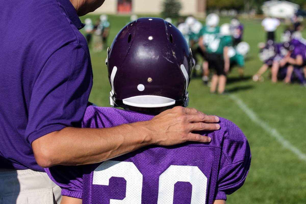
In a recent study in the Journal of Athletic Training, Schmidt et al. reported that youth tackle football players and parents do not accurately estimate the frequency, severity, or location of head impacts experienced during both practices and games. The findings revealed that players and their parents:
“Overestimated the head-impact frequency in practices but underestimated the frequency in games;
Overestimated head-impact severity, particularly in games;
Underestimated the number of head impacts to the top of the head, particularly during practices.”
One noteworthy finding was the underestimation of hits to the top of the head, as these can do the most damage due to the spine’s flexed position. Altogether, these results suggest that players and their parents have minimal understanding of various aspects of head impacts during tackle football. It is essential to understand the facts and data behind the game in the future to ensure the safety of players.
This study enlisted 16 players and 23 parents from four different recreational teams. Before the start of the season, players and parents completed a “head-impact estimation tool.” This survey consisted of 10 questions: three regarding frequency, three related to severity, and four related to location. Frequency was defined as the number of hits to the head; severity was defined as the acceleration of impact; and location was defined as the area of the head hit. Players were equipped with Smart Impact Monitors (SIM-G) to assess head-impact biomechanical data before every practice and game.
The study speculated that media, personal experience, and observation may all play a role in players’ and parents’ misconceptions about various aspects of head impacts. Television has been highly influential in people’s general understanding of football, as big tackles and hits are usually replayed multiple times.
Diagnostics
Analyzing auditory processing ability in the brain reveals history of subconcussive head impacts

Researchers Nina Kraus et al. at Northwestern University recently published a study in Exercise, Sport, and Movement analyzing the impact of subconcussive head impacts on a measure of the brain's auditory processing ability. They studied the auditory outcome measure FFR-F0, which measures the ability to hear and track another person's voice in a noisy environment. Previous research has found diminished FFR-F0 in individuals with an "active concussion" and those with a history of concussion.
In this study, Kraus et al. found that male contact sport players had reliably lower auditory processing results than their noncontact counterparts, even when controlling for past concussions. Kraus et al. raise concerns about the long-term impacts of contact sports. While the differences between the contact and noncontact groups were relatively small, the authors note that "college undergraduate athletes are, relative to professional athletes, at the beginning of their careers. If an effect is observable at this stage, it may be a figurative canary in a coal mine."
This study of NCAA Division I student-athletes found a slight correlation between the number of years in a contact sport and further decreased auditory processing results, especially for football and soccer players. This finding, and most of the other study findings, was found only in male athletes.
No reliable effect of contact vs. noncontact sports was found for female athletes, who also had higher FFR-F0 measures overall. These higher FFR-F0 measures imply the female brain has more auditory processing reserves and is more resilient to head trauma, which may explain why females report greater symptom severity after a concussion: females may need to sustain more damage to result in a concussion diagnosis, resulting in more symptoms.
The study also highlights the potential future use of the frequency-following response (FFR) measure "in the monitoring and early detection of subconcussive injuries that might lead to future cognitive difficulties and brain degeneration."
The participants included 709 division I college student-athletes, screened for normal hearing, and divided into prevailing classifications of contact (including limited-contact sports such as baseball) and noncontact sports. Participants were evaluated with the frequency-following response (FFR) to speech measure. FFR constructs a waveform based on the neural activity in a brain that is synchronized with a soundwave a subject is hearing. The study then evaluated the amplitude (size) of the fundamental frequency of the presented noise within the FFR of each subject. Nina Kraus and Travis White-Schwoch define the fundamental frequency (F0) as "the lowest dominating frequency of a sound that conveys the perception of pitch… The F0 is a crucial cue for picking a sound out from the din and tracking it." This FFR-F0 auditory outcome measure is a valuable tool to assess and monitor concussions and sub-concussions, as it doesn't rely on self-reported symptoms and is an objective marker that requires no input from the subject.
Therapies Currently Available
Brain fitness program improves symptoms in post-concussion syndrome patients
A study in the Journal of Alzheimer’s Disease Reports by Dr. Majid Fotuhi et al. explores a brain fitness program (BFP) that shows promise for people with post-concussion syndrome (PCS), attention-deficit/hyperactive disorder (ADHD), and memory loss. This non-pharmaceutical intervention provides an alternative treatment for people struggling with cognitive impairment and mood and sleep disturbances.
The 12-week program is aimed to promote neuroplasticity (the ability of the brain to grow and reorganize after injury) by providing patients with cognitive training, lifestyle coaching, and neurofeedback twice weekly. The researchers from George Washington University studied 223 child and adult patients enrolled in the BFP at the NeuroGrow Brain Fitness Center, an outpatient neurology practice in McLean, VA.
Fotuhi et al. found that the BFP cognitive training and coaching strategies and EEG neurofeedback helped improve brain health and cognitive function. Patients from all groups showed statistically significant improvements in cognitive scores, verbal memory, complex attention, processing speed, and executive functioning, with the exception of ADHD patients’ verbal memory scores. The greatest improvement was seen in patients struggling with PCS. Furthermore, 60-90% of patients reported a reduction in neurocognitive and neurobehavioral symptoms.
Patients between the ages of 7-80 were included in the study: 88 with PCS, 71 with ADHD, and 64 with memory loss. Prior to treatment, participants had quantitative electroencephalography (EEG) readings and neurocognitive testing, which included: verbal memory, complex attention, processing speed, and executive functioning. They also went through a comprehensive medical evaluation which looked at vitamin deficiencies and thyroid levels. In addition, patients completed questionnaires that assessed sleep, exercise, diet, and mood.
The BFP involves working one on one with brain coaches. Brain coaches provide individualized care and closely monitor patients’ progress. Implementing the Mediterranean diet, exercise plans, sleep schedules, calming strategies, and brain games are just a few of the strategies used during the program. Dr. Majid Fotuhi et al. theorize that these strategies may optimize glucose metabolism in the brain, improve cerebral blood flow, reduce inflammation, and increase brain-derived neurotrophic factor (BDNF) levels.
Overall, this research highlights the importance of multimodal interventions and individualized care. This program was shown to improve brain fitness and quality of life in people of all ages. The reduction in symptoms and improved cognition bring hope to others struggling with minor cognitive impairment.
Mental Health
Distinct connectivity patterns in TBI-associated depression suggest it is its own disorder, “TBI affective syndrome.” TMS is a potential treatment

A study found neurophysiological evidence suggesting TBI-associated depression is a distinct disorder from non-TBI depression, proposing the name “TBI affective syndrome.” Using resting-state functional connectivity MRI, the researchers found different patterns of connectivity between TBI-associated depression and non-TBI depression in the default attention network (DAN), default mode network (DMN), and the subgenual anterior cingulate cortex (sgACC).
The researchers say their study “suggests the need for different treatment approaches that target the specific pattern of disconnectivity seen in these patients.” Published in Science Translational Medicine, Shan H. Siddiqi et al. conclude, “...our results suggest that depressive symptoms after brain injury may represent a physiologically distinct ‘TBI affective syndrome’ that is fundamentally distinct from other forms of depression.”
In a Brigham and Women’s Hospital press release, Dr. Siddiqui notes that TBI-associated depression responds poorly to conventional antidepressants. The current study was predated by a pilot trial that found transcranial magnetic stimulation (TMS) effective for TBI-related depression. Unusual brain mapping findings in the pilot trial led to the current study identifying TBI affective syndrome. Several of the study authors will launch a multicenter trial to study the use of TMS to treat TBI-related depression. Study co-author Dr. Rajendra Morey says, “We hope our discovery guides a precision medicine approach to managing depression and mild TBI, and perhaps even intervene in neuro-vulnerable trauma survivors before the onset of chronic symptoms.” We recommend this Stat News article for further information.
Dr. Shan H. Siddiqi and colleagues at Harvard Medical School and Brigham and Women’s Hospital used a retrospective cross-sectional design to analyze data from five cohorts for a total of 273 participants. Participants formed four groups: “TBI without depression, TBI with depression, depression without TBI, or none of these disorders.” The researchers compared brain connectivity profiles across different groups of patients in order to determine whether TBI-associated depression is separate from non-TBI depression (MDD). They measured these connectivity profiles by utilizing resting-state functional connectivity MRI and a specific algorithm to create a map of each individual’s brain. Siddiqi et al. hoped these fMRI scans and maps would indicate significant differences in connectivity of the circuits of interest between TBI-associated depression and non-TBI depression groups, which would indicate different pathophysiology and corroborate findings that pharmacotherapy for non-TBI depression does not treat TBI-associated depression.
The study found that “TBI-associated depression was independently associated with decreased DAN–subgenual cingulate connectivity, increased DAN-DMN connectivity, and the combined effect of both.” Non-TBI depression was also associated with abnormal connectivity in these circuits but in the direction opposite of TBI-associated depression. On a generalized level, the DAN, DMN, and sgACC involve attention, reflection, and emotional behavior, respectively, and have been consistently implicated in depression pathophysiology. Because these circuits interact differently in these two types of depression, the pathophysiology likely differs, which supports the researchers’ proposal of a separate diagnosis for TBI-associated depression: TBI affective syndrome.
The retrospective cross-sectional nature of this study might have confounded the researchers’ results, even after they controlled for the effects of the cohort and replicated their results in another cohort; so, the researchers assert “...these retrospective results should be considered preliminary, and prospective studies are needed to better characterize this relationship.”
Women’s Health
We have two synopses this week: CTE and women's soccer
The world's first case of CTE in a former professional female athlete

Australian Sports Brain Bank researchers have reported the world's first chronic traumatic encephalopathy (CTE) diagnosis in a former professional female athlete. There have been several post-mortem CTE diagnoses in women, but this is the first in a professional female athlete. Heather Anderson, a 28-year-old Australian Football League for Women (AFLW) premiership player, died unexpectedly, and Catherine M. Suter et al. diagnosed Anderson with low-stage CTE. Their finding was published in Acta Neuropathologica.
An article in The Conversation suggests that finding CTE in Anderson's brain leads to questions about how a lifetime in contact sport may have contributed to her death. CTE has been increasingly associated with several contact sports, such as football, rugby, boxing, soccer, and martial arts. These sports have been dominated by male athletes, which accounts for the "male bias in CTE prevalence." Australian women and girls have increasingly sought to participate in football and contact sport; one million women and girls participated in contact sports in Australia in 2022, possibly increasing the risk for repetitive head trauma in this female population. A recent study found that lifetime exposure to cumulative force of repeated head impacts is strongly associated with CTE.
CTE, which can "only be definitely diagnosed after death," is a degenerative brain disease for which the only known risk factors are "prior exposure to traumatic brain injury (TBI; concussive and subconcussive). According to the Concussion Legacy Foundation, CTE symptoms during life may include mood and behavioral dysregulation and "progressive disorders of thinking and memory."
Anderson started contact sports early, joining a rugby team at just five years old, before going on to play Australian Rules football and eventually being drafted into the AFLW in 2017. Although she is the first professional female athlete diagnosed with CTE, she will certainly not be the last as more research progresses. This finding of CTE in an Australian former female professional athlete also comes shortly before the "inquiry into concussions and repeated head trauma in contact sports" ordered by the Australian Senate and due to be reported on September 5.
Women’s World Cup Soccer brings focus on concussions; safety recommendations

As the 2023 Fifa Women’s World Cup unfolds, media attention is brought to the risk of concussion for soccer players, particularly women, who are more vulnerable to this injury than men. According to an article by Shreva Mcleod and Kerry Peek in The Conversation, “for every 1,000 hours of playing or practicing soccer, there are about 1.5 concussions for women compared with 1.0 for men,” which may be due to factors such as a difference in neck strength, hormones, and lack of header-specific training.
According to this article, women who play soccer generally have weaker neck muscles than men, which makes it more difficult to stabilize their head and brace for impact from the ball or another player. According to one study, “higher neck strength may lower head acceleration during purposeful heading in soccer.”
In another study, adolescent soccer players who did not complete a program of neuromuscular neck exercises had a higher incidence of concussive events and were more likely to report a concussion than those who completed neuromuscular neck exercises.
Some hormones may be protective, such as estrogen and progesterone. While there is limited research, “Half of concussions also take place in the part of the menstrual cycle known as the ‘luteal phase,’” a week-long period in which estrogen and progesterone hormone levels are declining. However, the article states that, “the research is too limited to speculate further on the role of sex hormones.”
Finally, one last factor outlined by the article suggests that women are less likely than men to be trained to head the ball properly. A study found that high school girls closed their eyes when heading a ball at a higher rate than boys (90.6% vs 79%). Training such as “ocular motor and visual conditioning” could help players better prepare for contact with the ball and increase awareness of other players nearby, potentially reducing head-to-head contact.
The article also outlines possible guidelines to help reduce the risk of concussions.
To reduce the number of balls in the air and players on the field, thus potentially reducing the number of headers and collisions:
Reduce the number of players (for example, in training) to 5 players per side, or seven players per side
Utilize smaller goals in training
To reduce the force of balls that are headed:
Reduce the distance of balls played in the air. For example, the goalkeeper passes the ball to the defenders rather than making a long kick toward center field.
Do short corner kicks to nearby players to reduce the chance of header battles near the goal
Engage in exercises to help strengthen and stabilize muscles around the neck.
Give red cards (sending players off the pitch) for deliberate head contact.
Lastly, while research on women is lacking, recent studies have found that male soccer players at the elite (but not professional) level are at “significantly increased risk of neurodegenerative disease.” This increased risk appears to be position-specific (outfielders are more at risk) and may be due to “repetitive mild head trauma sustained through heading the ball and concussions.”